Table Of Content

During the catagen phase, hair growth ceases and the hair follicle shrinks. During a resting period, known as the telogen phase, the inferior portion of the follicle is missing. This phase leads back to the anagen phase and the follicle will regenerate to its full extent (Standring, 2016). The hair follicles in your skin contain living cells to allow your hair to grow.
Homology modelling of melatonin receptor 1A 3D structures and molecular docking

The hair strands were oriented in the X-ray diffractometer with their long axis along qz. The 2-dimensional data were integrated and converted into line scans and fit for a quantitative analysis. Another function of hair follicle is giving color to your hair. Hair, like your skin, gets its color from a pigment called melanin [6]. There are two types of melanin - eumelanin and pheomelanin.
What causes increased hair loss?
Your hair follicles are responsible for growing hair, which happens in cycles of three distinct phases. It can be due to drugs, diet, hormone imbalances, altered mitotic activity, growth cycle abnormalities, among others. A thorough history, physical exam, hair pull test, daily hair counts, part width, clip tests to examine the hair shaft, hair growth windows, and hair pluck, and trichograms can all be used to diagnose hair disease. Scalp biopsies, hormone studies, and a potassium hydroxide examination for fungi may also need to be performed in certain cases.
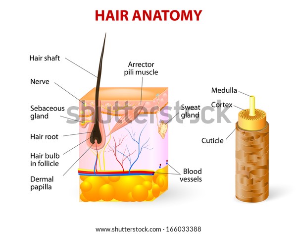
How Do Hair Follicles Function?
The identical X-ray signals indicate that these products do not have an effect on the molecular structure of keratin and membranes deep inside the hair (within the resolution of our experiment). As expected, the identical twin pair shows almost identical hair structures whereas the fraternal pair exhibits distinct differences. Offspring receive half of their chromosomes from each parent, thus the genetic similarity between the parent and child pair is roughly the same as fraternal twins (Creasy et al., 2013).
Telogen effluvium
Fig. 4. Histopathologic features of hair follicles in normal controls,... - ResearchGate
Fig. 4. Histopathologic features of hair follicles in normal controls,....
Posted: Thu, 05 Sep 2019 16:31:57 GMT [source]
During the development of bullous peg (stages 5–8), the hair bulb and the main cell layers of the mature hair follicle are also formed [2–4, 6]. Hair grows and is eventually shed and replaced by new hair. The first is the anagen phase, during which cells divide rapidly at the root of the hair, pushing the hair shaft up and out. The length of this phase is measured in years, typically from 2 to 7 years. The catagen phase lasts only 2 to 3 weeks, and marks a transition from the hair follicle’s active growth.
The packing of these fibrils by bundling into macro-fibrils is characterized by X-ray diffraction pattern by three equatorial spots located at about 90, 45 and 27 Å (Busson, Engstrom & Doucet, 1999). The corresponding signals are observed in the 2-dimensional data in Fig. The exact position of the features is, however, best determined in small angle diffraction experiments (SAXS), which offer a drastically improved resolution, and will be shown below. We note that the axial packing of coiled-coils within keratin filaments in hair gives rise to a number of fine arcs along the meridian (z). While the features observed in scattering experiments are well known, the molecular architecture of the intermediate filaments is still under discussion (Rafik, Doucet & Briki, 2004). Supercoiled coiled-coils or models that involve straight dimers with different numbers of coils are being discussed.
The new hair shaft appears on the skin's surface to mark the metanagen phase. In darkly pigmented individuals, melanin can be found in abundance within the melanophages of the dermal papilla. The hair matrix is the actively growing portion of the follicle consisting of a collection of epidermal cells that rapidly divide, move upward, and give rise to the hair shaft and the internal root sheath. In histological sections, hair follicles may have different appearances. This is because the hair undergoes cycles of growth and loss. In the anagen phase, the hair is actively growing, and the follicle is at is maximal extent of development.
The hair also plays important roles for the individual’s social and sexual interaction [1, 2]. This can occur on the entire scalp (alopecia totalis) or body (alopecia universalis) or may be localized to specific areas (alopecia areata). Alopecia areata can progress to alopecia totalis and often begins acutely and spreads gradually over the course of weeks to months. Alopecia is an autoimmune disease mediated by T lymphocytes and other immune cells. During periods of stress, more hair enters the telogen phase and begins to fall out.
Hair texture (straight, curly) is determined by the shape and structure of the cortex, and to the extent that it is present, the medulla. The shape and structure of these layers are, in turn, determined by the shape of the hair follicle. Hair growth begins with the production of keratinocytes by the basal cells of the hair bulb.
In predisposed individuals, the terminal hairs on the adult scalp can undergo involutional miniaturization (become vellus). The upper part named acroinfundibulum, the keratinization of epithelium turns into the “epidermal mode”, with formation of stratum granulosum and stratum corneum like a similar manner to epidermis [1, 14, 16]. Several molecular pathways, growth factors, proteins and genes play substantial roles for the development of the hair follicle. Canonical (β-catenin dependent) WNT (wingless-type integration site) signals are candidates for the initial dermal message, and it is believed that they precede other activators and regulators of appendage development.
The two signals present in all individuals in the equatorial plane (q‖) correspond to the distance between two coiled coils of 9.5 Å and between two lipid tails in the cell membrane cortex of 4.3 Å. The common meridional signal along the long axis of the hair (qz) at 5 Å corresponds corresponds to the α-helices twisting around each other within coiled-coils. The majority of hair fibre is the cortex which contains spindle shaped cells that lie parallel along the fibre axis. These cortical cells were found to be approximately 1–6 µm in diameter and 50–100 µm in length (Randebrook, 1964).
However, it’s best to speak to a dermatologist if you think you have telogen effluvium, because they’ll need to rule out other causes. According to the U.S National Library of Medicine, 50 million men and 30 million women are affected by androgenetic alopecia. An abundance of eumelanin makes hair black, a moderate amount of eumelanin makes hair brown, and very little eumelanin makes hair blonde. Your genes determine whether you have eumelanin or pheomelanin, as well as how much of each pigment you have.
Lineage studies have proven that bulge cells are multipotent and that their progeny generate the new lower anagen hair follicle [21]. One of the most distinguishing features of stem cells is their slow-cycling nature, presumably to conserve their proliferative potential and to minimize DNA errors that could occur during replication. On entering the hair bulb matrix, they proliferate and undergo terminal differentiation to form the hair shaft and inner root sheath. They also migrate distally to form sebaceous glands and to proliferate in response to wounding [16, 20, 22].
It is interesting to note that differences are observed for the fraternal twins in Fig. This finding is in agreement with the expectation that individuals with similar genetics would share similar physical traits such as hair structure. Identical or monozygotic twins originate from one zygote during embryonic development, and they share 100% of their genetic material.
Hair that does grow is thinner and more fragile hair than normal. This can lead to hair loss at the site of your injured skin. Differences in the X-ray data between individuals were observed in the wide angle region (WAXS) of the 2-dimensional data in Fig.
Two articles had both hair follicle findings and litter size. Hair follicle, litter size, in vitro oocyte, and gene data performance traditional and network meta-analysis. Traditional meta-analysis of follicle density, cashmere fibre diameter, and yield. Traditional meta-analysis of melatonin’s effect on cashmere fibre length. Traditional meta-analysis of melatonin’s effect on goat litter size.


No comments:
Post a Comment